
These HSC Biology Module 6 notes cover Genetic Change in the NSW Year 12 course: how genetic information changes, how those changes spread through groups of living things, and how people use and alter living things. Every term is explained from the ground up.
Part of our HSC Biology notes. These notes follow the Biology Stage 6 Syllabus (2017) for the 2026 and 2027 HSC. The new syllabus is first examined in 2028.
The Working Scientifically skills for Module 6 near the end of this page cover investigation design, data and evidence. The HSC Biology past-papers guide provides official papers and marking guidelines.
Key Takeaways
- Only a mutation (a lasting change in DNA) creates a new allele, a version of a gene. Sexual reproduction only reshuffles existing alleles.
- A change in DNA, a change in the organism and a change in the gene pool (all the alleles of a population) are separate steps; one does not guarantee the next.
- Biotechnology uses living things to make products or do jobs, from bread-making and selective breeding to reading, copying and editing DNA.
- Judge each technology on evidence: how well it works, how it affects the variety of life (biodiversity), and its social, economic and cultural context.
This module is about how genetic information changes, and how people now change it on purpose. Genetic change happens at three levels, one after another:
- The DNA changes. A letter in the genetic code is swapped, lost or added.
- The organism may change. The changed DNA might alter how the body works. Often it alters nothing you can see.
- The population may change. If the change is passed to offspring, it can become more or less common over generations.
Each step can happen without the next one. For example, a change in one part of a person’s body is never passed to their children. The words below are used throughout the module.
Start here: the basic words of genetics
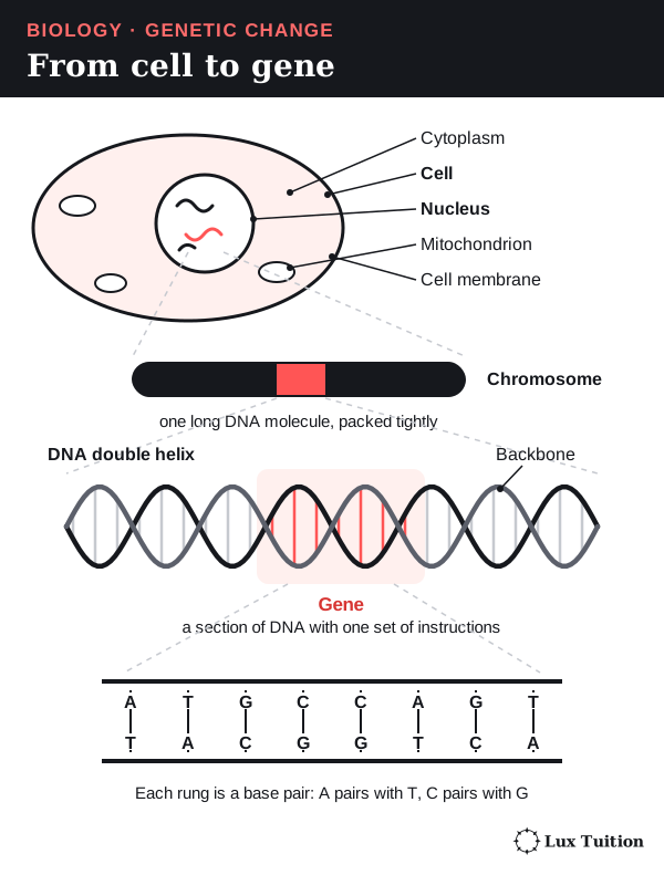
Every living thing is made of cells, the smallest units that can carry out life’s processes. A group of similar cells working together is a tissue. Inside a cell is a jelly-like fluid, the cytoplasm. In plants, animals and fungi, most cells also have a nucleus, a compartment that holds the genetic information, and mitochondria, small structures that release energy from food.
The genetic information is stored in DNA (deoxyribonucleic acid), a long molecule shaped like a twisted ladder, called a double helix. DNA is built from units called nucleotides. Each nucleotide has a sugar, a phosphate and one of four bases: A (adenine), T (thymine), C (cytosine) or G (guanine).
The sugars and phosphates form the two sides of the ladder, called the backbone. The bases pair up to form the rungs. A always pairs with T, and C always pairs with G. This rule is complementary base pairing, and it means one strand can always be used to rebuild the other.
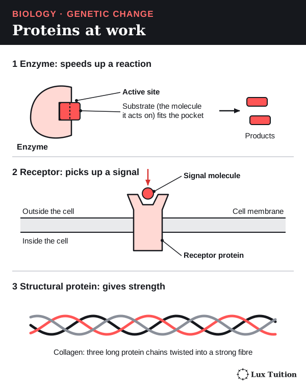
The order of the bases is the DNA sequence. A gene is a section of DNA that carries the instructions for one product, usually a protein. Proteins do most of the work in a cell:
- Enzymes speed up chemical reactions. Each has an active site, a pocket that fits the molecule it acts on.
- Receptors sit in the cell membrane (the thin outer layer of a cell) and pick up chemical signals.
- Structural proteins, such as collagen, are long fibres that give tissues strength.
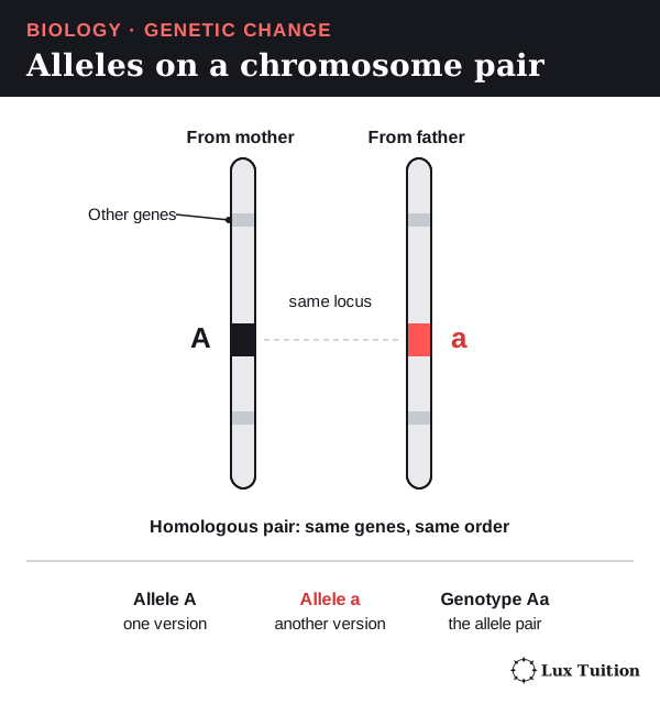
A chromosome is one very long DNA molecule, packed tightly with proteins. Humans have 46 chromosomes in most body cells, in 23 pairs. The two chromosomes in a pair are homologous chromosomes: they carry the same genes in the same order, one from the mother and one from the father.
A cell with chromosome pairs is diploid (2n). A gamete (egg or sperm) carries only one chromosome from each pair, so it is haploid (n).
An allele is one version of a gene. For example, a gene for flower colour might have a purple allele and a white allele. The position of a gene on a chromosome is its locus.
Because chromosomes come in pairs, a diploid organism carries two alleles of each gene. This allele pair is its genotype (for example Aa). Its observable characteristics are its phenotype.
A dominant allele shows its effect even when only one copy is present. A recessive allele shows its effect only when both copies are recessive.
A mutation is a lasting change in the DNA sequence, and it is the only way a brand-new allele appears. A population is a group of the same species living and breeding in one area. All the alleles carried by its breeding members make up its gene pool.
Keep four ideas separate through the whole module: making a new allele, reshuffling existing alleles, copying DNA, and changing how common alleles are in a population.
How does mutation introduce new alleles into a population?
How do mutagens damage DNA and cause mutations?
electromagnetic radiation sources
chemicals
naturally occurring mutagens
What a mutation is
Some mutations change a single base. Others, called chromosome mutations, change the arrangement or number of whole chromosomes.
Mutations are random with respect to need: an organism does not mutate because it “needs” a new trait. A harmful agent can make mutations more frequent, but it cannot aim them at a useful result.
Before a cell divides, it copies all of its DNA. This is DNA replication. The enzyme DNA polymerase builds each new strand by reading an old strand and adding the complementary base each time.
Mutagens: what increases the mutation rate
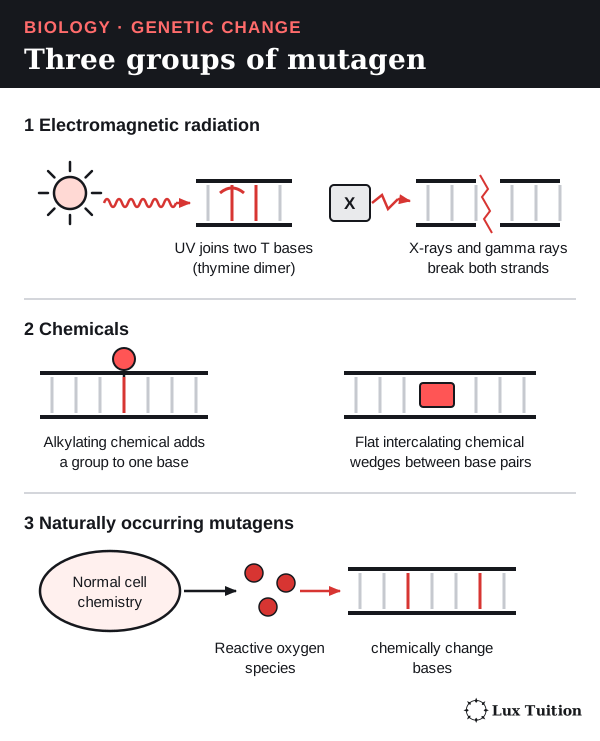
A mutagen is anything that makes mutations more likely. The syllabus names three groups. Where an agent comes from and how it works are separate questions: sunlight is natural, but it is also radiation. Not every chemical is a mutagen, and some carcinogens promote cancer without directly changing DNA.
1. Electromagnetic radiation.
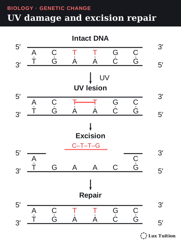
- Ultraviolet (UV) light, including sunlight, can join two neighbouring pyrimidine bases (C and T in DNA) on the same strand. Two joined thymines form a thymine dimer, which bends the DNA, so polymerase may copy the wrong base opposite it. UV only reaches the body’s surface, not the eggs or sperm inside.
- X-rays and gamma rays are ionising radiation: they carry enough energy to knock electrons off atoms. They can break one or both DNA strands, and can also make damaging reactive molecules from water. If the broken ends are rejoined wrongly, chromosome pieces can be lost or rearranged.
2. Chemicals.
- Alkylating chemicals attach small chemical groups to bases. A changed base may pair with the wrong partner during replication, causing a substitution (one base swapped for another).
- Intercalating chemicals are flat molecules that slip between the rungs of the DNA ladder. Polymerase may then add or skip a base, causing an insertion (extra bases) or a deletion (missing bases).
3. Naturally occurring mutagens.
- Reactive oxygen species are reactive by-products of the cell’s own chemistry. They can change bases so that they pair wrongly. Being natural does not make them harmless.
Mutations also happen with no mutagen at all. These spontaneous mutations include rare copying errors, the loss of a base, and a base changing by itself (for example deamination, cytosine turning into a different base, uracil, which pairs wrongly).
Risk depends on the dose, the type of cell and how well the cell repairs damage. In an exam, explain the whole chain: mutagen or copying error → DNA damage → failed or wrong repair → permanent change copied into new cells. NHGRI mutagen definition.
Damage is not yet a mutation
Damage only becomes a mutation if it is copied or repaired wrongly. Cells have three main defences:
- Proofreading: DNA polymerase checks each base it adds and removes most mistakes straight away.
- Mismatch repair: enzymes find wrongly paired bases left after copying and fix them.
- Nucleotide excision repair: enzymes cut out a short damaged stretch of one strand. DNA polymerase rebuilds it, using the undamaged strand as a template. DNA ligase then seals the gap in the backbone.
If damage escapes these defences, the wrong sequence is copied into new cells and becomes permanent. DNA repair overview.
How do point mutations and frameshift mutations differ?
point mutation
Start here: how a gene makes a protein
A gene is used in two stages.
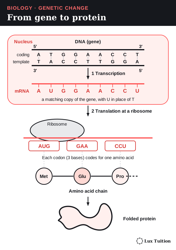
- Transcription. In the nucleus, the gene’s DNA is copied into a matching molecule called messenger RNA (mRNA). RNA uses the base U (uracil) in place of T.
- Translation. The mRNA travels to a ribosome, the cell’s protein-building machine. The ribosome reads the mRNA three bases at a time. Each group of three is a codon. Most codons stand for one amino acid, the building block of proteins; a few are stop codons, which end the protein.
The amino acids are joined in the order of the codons, and the chain folds into a protein. The reading frame is the way the bases are grouped into threes, counting from the first codon.
Each strand of DNA has two different ends, called 5′ and 3′ (“five prime” and “three prime”). The two strands of the helix run in opposite directions, which is called antiparallel: one runs 5′ → 3′ and its partner runs 3′ → 5′.
The template strand is the one read to build the mRNA. The other strand, the coding strand, has the same sequence as the mRNA (with T instead of U). The mRNA is complementary and antiparallel to its template strand. Mixing up these strands gives the wrong amino acids in an answer.
Types of point mutation
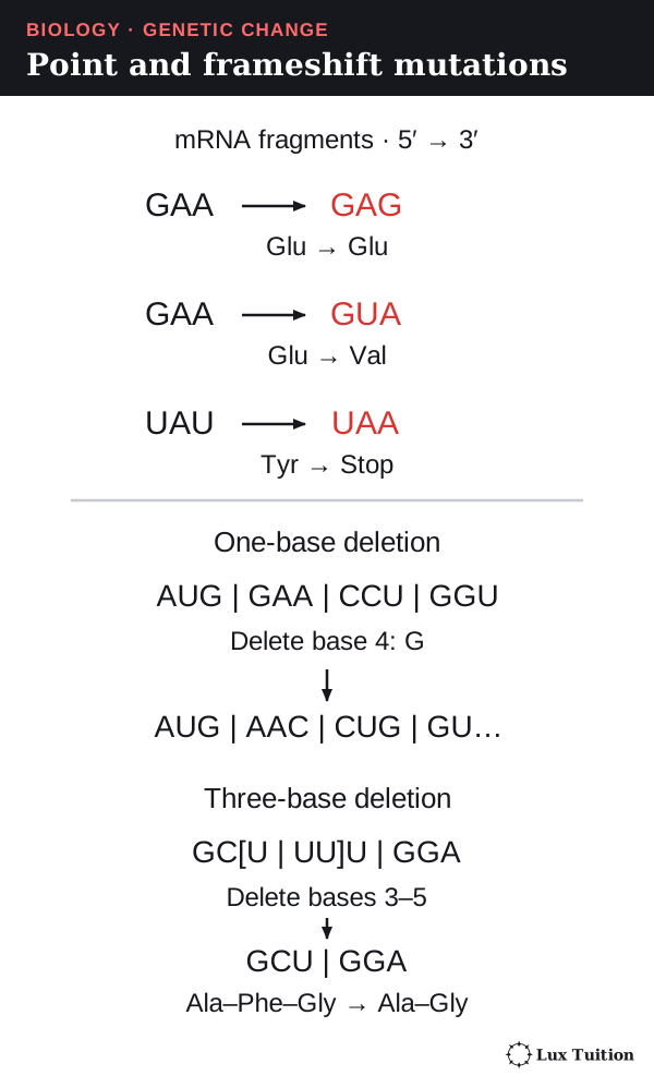
A point mutation changes a single base pair. It can be a substitution, which keeps the DNA the same length, or an insertion or deletion of bases. Some textbooks use “point mutation” mainly for substitutions, so name the exact change (for example “a substitution of A for G”). NHGRI point mutation.
What each change does to the protein
The examples use mRNA, written 5′ to 3′ (the direction the ribosome reads it).
| Change | Example (mRNA) | What happens to the protein |
|---|---|---|
| Silent (synonymous) substitution | GAA → GAG. Both code for glutamate. | Same amino acid. (It can still affect splicing, the cutting and joining of RNA, or expression, how much of a gene’s product is made.) |
| Missense substitution | GAA → GUA. Glutamate becomes valine. | One amino acid changes. The effect depends on where it is and how different the new one is. |
| Nonsense substitution | UAU (tyrosine) → UAA, a stop codon. | Translation stops early; the short protein usually cannot work, and premature stops can reduce mRNA stability. |
| Frameshift (insertion or deletion of 1 or 2) | AUG–GAA–CCU–GGU. Delete the G after AUG: AUG–AAC–CUG–GU… | Every codon after the change is regrouped, often reaching an early stop codon. |
| In-frame insertion or deletion (3, 6, 9…) | Three bases added or removed. | The frame after the change is kept, but nearby amino acids are added, lost or altered. The protein can still fail. |
Think of the reading frame like a sentence of three-letter words: THE CAT SAT. Delete the C and the reader keeps counting in threes: THE ATS AT… (DNA has no spaces: the cell counts in threes from the start codon.)
A three-base deletion does not always remove one whole codon. Take GCU–UUU–GGA (alanine–phenylalanine–glycine). Delete the third base of GCU and the first two bases of UUU.
The surviving U supplies the third base, so the rest regroups as GCU–GGA. One amino acid is lost, and the codons after it are unchanged. Always regroup into threes before deciding the effect.
The effect on the organism also depends on where the change falls, what the protein does, whether the allele is dominant, other genes and the environment. A small change in an enzyme’s active site can matter more than a larger change elsewhere. MedlinePlus variant types.
How do chromosomal mutations change chromosome structure and number?
chromosomal mutation
Start here: mitosis and meiosis
Cells make new cells by dividing. In animals, there are two ways:
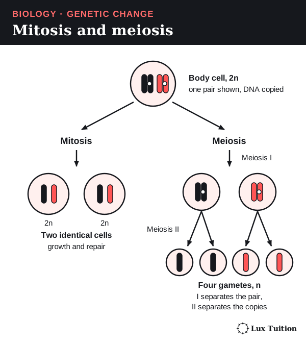
- Mitosis makes two identical cells, each with the same chromosome number as the parent cell (2n in a body cell). It is used for growth and repair.
- Meiosis makes gametes. It has two divisions, meiosis I and meiosis II, and produces four cells with half the chromosome number (n). In meiosis I the homologous pairs separate; in meiosis II the two copies of each chromosome separate.
Plants differ slightly: in plants, meiosis makes spores, and the gametes are later made from them by mitosis.
DNA is copied once, before mitosis or before meiosis I, and not again between the two meiotic divisions. Each chromosome is then made of two identical copies. These copies, called sister chromatids, stay joined at a pinch point called the centromere.
Changes to chromosome structure
A chromosome mutation changes a large piece of a chromosome, so it can affect many genes at once. Two main causes are:
- Breakage and wrong rejoining, for example after ionising radiation.
- Unequal crossing over. During meiosis, matching chromosomes swap pieces (crossing over, explained under meiosis below). If they line up out of place, one gains an extra copy of a section and the other loses it.
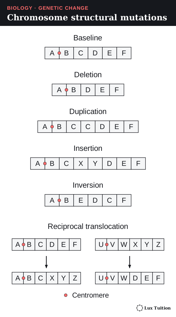
Each letter below stands for one section of a chromosome.
| Change | Example | Possible effect |
|---|---|---|
| Deletion | ABCDEF → ABDEF | Genes in C are lost, so less of their product may be made. |
| Duplication | ABCDEF → ABCCDEF | Extra gene copies can raise the amount of product (gene dosage); over time a spare copy can gain a new job. |
| Insertion | ABCDEF → ABCXYDEF | A section from elsewhere can break up a gene or its control region. |
| Inversion | ABCDEF → ABEDCF | Break points can split a gene, and pairing in meiosis can be upset. |
| Translocation | A section moves to a non-matching chromosome. | Break points can split or fuse genes; carriers can make gametes with extra or missing pieces. |
A balanced change has no overall gain or loss of DNA, but its break points can still disrupt genes. An unbalanced change alters the amount of DNA. MedlinePlus chromosome structure.
Changes to chromosome number
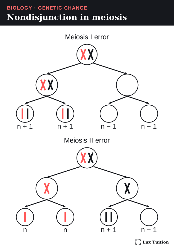
A change in number usually starts with nondisjunction: chromosomes fail to separate in meiosis. In meiosis I, a homologous pair fails to separate; in meiosis II, two sister chromatids fail to separate.
The gametes then have one chromosome too many (n + 1) or too few (n − 1). After fertilisation (the joining of two gametes) with a normal gamete, the offspring has:
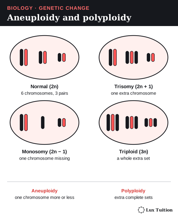
- trisomy (2n + 1), as in Down syndrome (trisomy 21), or
- monosomy (2n − 1).
An extra or missing single chromosome is called aneuploidy. Exams often test the counts (assuming one affected pair and otherwise normal segregation):
- Error in meiosis I: two products are n + 1 and two are n − 1.
- Error in one meiosis II division: two products are normal, one is n + 1 and one is n − 1.
In human females only one of the four products becomes a working egg. Nondisjunction in mitosis after fertilisation instead makes a mosaic: a mix of cells with different chromosome numbers.
Polyploidy is different: the cell has whole extra sets of chromosomes (3n, 4n), not one extra chromosome. It can happen when a cell copies its chromosomes but fails to divide, or when two unreduced (2n) gametes join. It is common in plants; wheat is polyploid.
A karyotype is a photograph of a cell’s chromosomes, lined up in pairs. It shows chromosome number and large structural changes, but not single-base changes, so a normal karyotype does not rule out a genetic disorder.
The first 22 pairs are autosomes (non-sex chromosomes). The last pair is the sex chromosomes: XX is usually female and XY usually male. Read this pair separately from any number change. For example, a karyotype with three copies of chromosome 21 and XX shows a female with a chromosome number mutation (trisomy 21), not a deletion.
Size alone does not decide how serious a mutation is. A number change alters the dose of hundreds of genes, but a single substitution can be just as harmful if it disables an important protein. MedlinePlus chromosome number.
How do somatic and germ-line mutations differ?
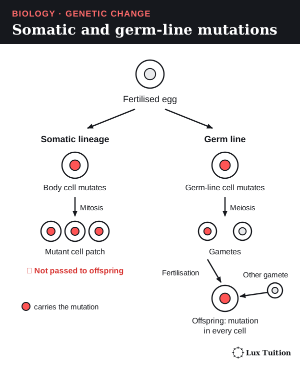
An animal body has two kinds of cell line. Somatic cells are all the body cells except those that make eggs and sperm. The germ line is the small set of cells that make gametes. Only the germ line is passed on to children.
Somatic mutations
A somatic mutation happens in a body cell. Then:
- The cell divides by mitosis, so every cell it produces carries the mutation.
- This forms a patch of mutated cells in an otherwise normal body.
- The patch may have no effect, may weaken a tissue, or may help cause cancer.
- Eggs and sperm do not come from that patch, so the mutation is not passed to offspring.
Cancer is uncontrolled cell division. A proto-oncogene promotes division when the body needs it; a mutation that switches it on permanently is like a stuck accelerator. A tumour-suppressor gene stops division or makes damaged cells die, so losing it removes the brakes.
Cancer usually needs several such mutations in one cell line. A somatic mutation can therefore seriously harm its carrier without being inherited by their children.
Germ-line mutations
A germ-line mutation happens in a gamete or a cell that will make gametes.
- Meiosis passes the mutation into some of the gametes.
- If one of those gametes is used in fertilisation, the offspring starts life with the mutation.
- Normally every cell of the offspring is copied from that first cell, so the mutation is in every cell, including its own germ line, and can be passed on again.
A mutation in the fertilised egg before its first division can enter all descendant cells. After division has begun, a mutation in one cell of the early embryo (the developing organism soon after fertilisation) may produce mosaicism, because only that cell’s descendants inherit it. A parent can also pass on a mutation present only in their germ line.
“Heritable” means the mutation can be passed on, not that it will be. The gamete might never be used, and the effect may depend on the other allele the child inherits.
The somatic/germ-line split is a simplified animal model. A plant has no germ line set aside early, so a mutation in a shoot can end up in flowers grown from that shoot. Plant shoot mutations can also pass through cuttings. MedlinePlus origins of variants.
Why do mutations in coding and non-coding DNA matter?
Only a small part of our DNA spells out proteins. Much of the rest controls when, where and how much each gene is used.
The parts of a gene
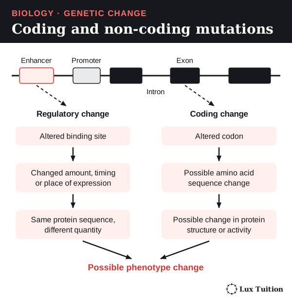
- Coding DNA is translated into a protein’s amino acid sequence.
- Non-coding DNA is everything not translated into protein. It includes:
- the promoter, the “start here” switch before a gene, where the machinery that makes mRNA attaches;
- enhancers, control regions that turn a gene up in certain tissues or at certain times. Proteins called transcription factors bind to promoters and enhancers to do this;
- introns, stretches inside a gene that are copied into RNA and then cut out. The pieces kept and joined are exons (exons can include untranslated regions), and the cutting and joining is splicing;
- untranslated regions, the ends of the mRNA that are kept but not read into amino acids;
- genes for functional RNAs, which work as RNA and are never made into protein.
Where a mutation lands changes what it does
- In coding DNA: a codon, the reading frame or a stop signal can change, so the protein itself may change.
- In a promoter or enhancer: the protein sequence may stay unchanged, but how much is made, when or in which tissue can change.
- At a splice site (the edge of an intron): part of an exon can be skipped or part of an intron kept, which can shift the reading frame.
- In a functional-RNA gene: that RNA’s own job can be affected.
Many non-coding mutations have no detectable effect. Location tells you what could happen; evidence shows what does.
Worked comparison. A regulatory mutation that cuts transcription of an enzyme gene gives less of a normal enzyme. A coding mutation can give a normal amount of a less active enzyme. Both slow the pathway, but by different causes. MedlinePlus non-coding DNA and non-coding variants and health.
How do mutation, meiosis and fertilisation create genetic variation?
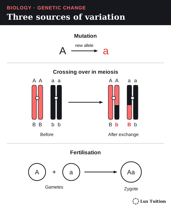
Brothers and sisters look different even though they have the same parents. Three processes explain this:
- Mutation creates new alleles.
- Meiosis reshuffles a parent’s existing alleles into new combinations in their gametes.
- Fertilisation combines one gamete from each parent.
Only mutation makes something genuinely new; the other two rearrange what already exists.
Meiosis: crossing over
Early in meiosis I (prophase I), the two chromosomes of each homologous pair line up side by side. Each has already been copied, so there are four chromatids. Chromatids from different chromosomes of the pair are non-sister chromatids.
In crossing over, two non-sister chromatids break at the same point and swap matching pieces. So a chromosome carrying A and B and its partner carrying a and b can produce chromatids carrying A b and a B. No new allele is made; existing alleles are recombined.
Meiosis: independent assortment
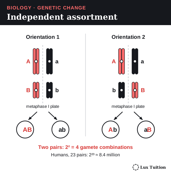
In metaphase I, the homologous pairs line up across the middle of the cell. Each pair lines up independently, so whether the mother’s or father’s chromosome faces a given side is random. Each gamete therefore gets a random mix of maternal and paternal chromosomes. This is independent assortment.
With n chromosome pairs there are 2n possible combinations. Humans have 23 pairs, so 223 ≈ 8.4 million, before crossing over adds more. This count assumes independent sorting and counts whole chromosomes only.
Fertilisation
At fertilisation, each gamete brings one haploid set, so the new cell, the zygote, is diploid again. Which sperm meets which egg is chance, adding more variation.
Two Aa parents each make A and a gametes, so their children could be AA, Aa or aa. Fertilisation makes new genotype combinations, but it did not create A or a; only an earlier mutation did. A mutation that happens while gametes are being made is still mutation, not part of normal reshuffling.
How do mutation, gene flow and genetic drift change gene pools?
Start here: allele frequency
Allele frequency is how common one allele is in the gene pool, as a proportion of all copies of that gene. For a gene on an ordinary (non-sex) chromosome, each diploid individual carries two copies:
- f(A) = number of A alleles ÷ total number of alleles for that gene
- f(A) + f(a) = 1 when there are only two alleles.
When allele frequencies change over generations, the population is evolving. Four processes can change them: mutation, gene flow, genetic drift and natural selection.
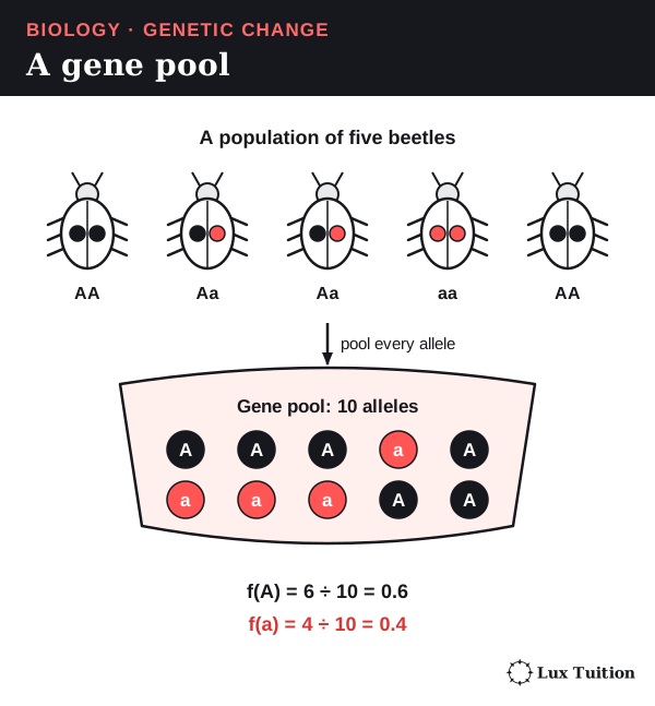
Mutation, gene flow and genetic drift
| Process | What happens | Effect on the gene pool |
|---|---|---|
| Mutation | A new allele appears in a gamete and is inherited. | Adds new variation, at first very rare. Even a helpful mutation is not guaranteed to spread. |
| Gene flow | Individuals or their gametes move between populations and breed. | Can bring in a missing allele and make connected populations more alike. A migrant who does not breed adds nothing. |
| Genetic drift | Allele frequencies change by chance, because only some individuals happen to breed. | Strongest in small populations. Can remove an allele or fix it (make it the only allele left), whatever its advantage. It often reduces variation within a population while increasing differences between isolated populations. |
Drawing ten marbles from a half-red, half-blue jar often gives six or seven of one colour by luck; a hundred draws land closer to half and half. That is why drift is strongest in small populations.
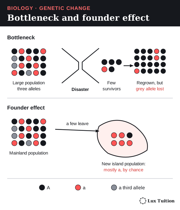
Two events make drift especially strong:
- A bottleneck: the population crashes (for example after a fire), leaving a few unrepresentative survivors.
- A founder effect: a few individuals start a new population, carrying only some of the original alleles.
If the population grows again, lost alleles do not return unless mutation or gene flow brings them back. NHGRI genetic drift.
Natural selection is different from drift. Individuals with certain heritable traits survive and reproduce more in a particular environment, so their alleles become more common. Spraying a pesticide favours insects that already carry a resistance allele; it does not make insects develop the mutation. Selection and drift can act together, so a frequency change alone does not show its cause.
Worked example: calculating allele frequency
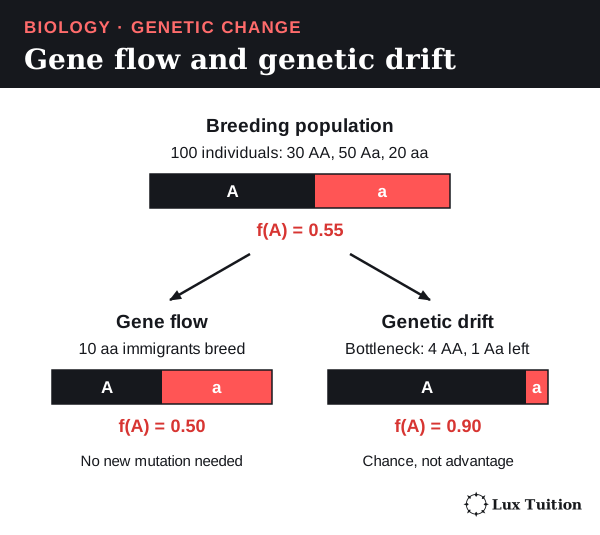
Step 1: count the alleles. A population has 100 diploid individuals: 30 AA, 50 Aa and 20 aa.
- A alleles = (2 × 30) + 50 = 110, out of 2 × 100 = 200
- f(A) = 110 ÷ 200 = 0.55, so f(a) = 0.45
Step 2: gene flow. Ten aa individuals join and breed. A is still 110, out of 220: f(A) = 0.50. No mutation was needed.
Step 3: a bottleneck instead. A disaster leaves four AA and one Aa: 9 A out of 10 alleles, so f(A) = 0.90. A became common by luck, not because it helped survival.
The numbers are invented and assume every counted individual breeds with equal reproductive contributions; real transmission can differ. Population genetics and allele counts.
How do genetic techniques affect Earth’s biodiversity?
How has biotechnology changed from fermentation to genetic engineering?
Biotechnology is using living things, their cells or their molecules to make useful products or do useful jobs. It is much older than modern DNA techniques.
Past: using organisms without understanding them
Microorganisms are living things too small to see without a microscope. Fermentation is a process in which microorganisms break down sugars without oxygen. Yeast, a single-celled fungus, is the classic example.
- In bread making, yeast releases carbon dioxide gas, which makes the dough rise.
- In brewing and wine making, it also produces ethanol (alcohol).
Selective breeding means choosing which plants or animals with useful traits are allowed to breed. Over many generations the chosen individuals had more offspring, so the alleles behind those traits became more common, long before DNA was discovered.
Present: working with DNA directly
Bacteria are single-celled microorganisms with no nucleus. They and yeast are the workhorses of modern biotechnology, which uses these tools:
- Large-scale culture: growing huge numbers of bacteria or yeast in tanks to make products.
- DNA sequencing: reading the order of bases in DNA.
- Polymerase chain reaction (PCR): making millions of copies of one chosen piece of DNA.
- Recombinant DNA: joining DNA from different sources, such as putting a human gene into a plasmid (a small ring of DNA) inside a bacterium.
- Genome editing: changing DNA at one chosen place in an organism, often with Cas9, an enzyme that cuts DNA at a chosen spot.
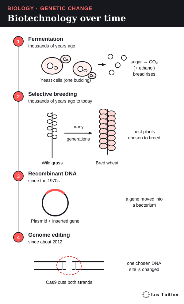
Genetic engineering means directly changing an organism’s genetic material with these tools. Selective breeding, by contrast, only changes which existing alleles are passed on.
The tools do different jobs. Sequencing only reads DNA and PCR only copies it; neither changes a living organism. A genome edit can create a new allele.
| Approach | How it works | Main advantage | Main limit |
|---|---|---|---|
| Selective breeding | Parents with useful traits are bred, and the best offspring chosen again. | Uses variation already in the population. | Unwanted alleles linked to a chosen trait can come along too. |
| Hybridisation | Two genetically different varieties are crossed. | Can combine traits; offspring may grow better than either parent (hybrid vigour). | Traits can depend on several genes and the environment; later generations vary; some cross-species hybrids are infertile. |
| Reproductive technologies | People help gametes meet or embryos develop. | More offspring from chosen parents, even across long distances. | No new DNA sequence is made; overusing a few parents narrows diversity. |
| Recombinant DNA | DNA pieces are joined and put into a host organism. | Moves one defined gene; can make a useful protein in bulk. | The gene may not be inherited or switched on correctly. |
| Genome editing | A chosen DNA site is changed, often with a guided enzyme. | Can change the organism’s own gene without adding another species’ gene. | Desired edits can have biological trade-offs; delivery, unintended changes and repair outcomes all need testing. |
The newest technique is not automatically the best.
What social and ethical issues arise in plant and animal biotechnology?
analysing the social implications and ethical uses of biotechnology, including plant and animal examples
Science can measure a benefit or a risk. Deciding whether it is acceptable also involves values. Ethics is the study of what is right, fair or important. A useful checklist:
- Benefit: who gains, and how much?
- Harm: who or what could be hurt, including animals and the environment?
- Informed choice: do people know enough to decide?
- Fairness: who can afford or access it?
- Stewardship: are we looking after the environment and future generations?
Two people can agree on the science and still disagree, because they weigh these points differently.
Plant example. A farmer may value reliable harvests and fewer pest losses, but must weigh seed prices, licence conditions and depending on one supplier. Consumers may care about price, nutrition, the environment or labelling. Neighbouring farmers may worry about pollen drifting onto their crops. Market access, separate supply chains and adequate storage also matter.
A higher-yielding crop does not by itself fix poverty or unequal food distribution. Patents and contracts are legal rules; they are not evidence that genetically modified seeds are sterile.
Animal example. A high-performing livestock breed can bring farm benefits, but an ethical evaluation also counts the welfare of donor animals, surrogate mothers (females who carry another animal’s embryo) and offspring. Cloning (making genetically identical copies) can cause failed pregnancies and developmental problems, and extreme production traits can compromise animal health and mobility. In research, the value of the question must justify using animals, and alternatives must be considered.
What are the future directions of biotechnology?
researching future directions of the use of biotechnology
Future research aims to make editing more accurate, deliver it to the right cells and regulate it well. Farm research looks for crops that resist disease or cope with drought. Conservation research combines frozen seeds, sperm and eggs with reproductive technologies to help endangered species.
These are directions, not promises. Complex traits such as drought tolerance involve many genes and the environment, so changing one sequence rarely solves the whole problem.
How could gene drives change a population?
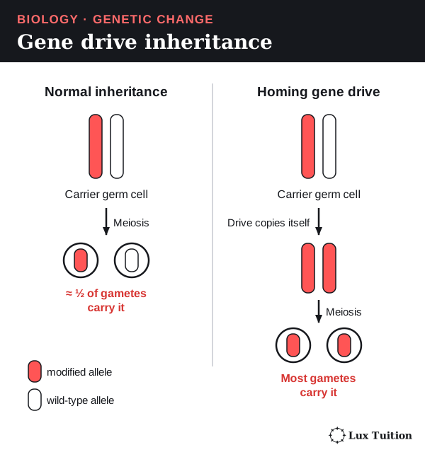
Normally, a parent with one copy of an allele passes it to about half of its offspring, as Mendel described. A gene drive is an engineered piece of DNA that breaks this rule, so it reaches far more than half of the offspring.
A homing gene drive works like this:
- The drive sits on one chromosome. It carries the gene for a DNA-cutting enzyme plus a short “guide” that steers the enzyme to the matching spot on the partner chromosome (this is the CRISPR–Cas9 system explained later on this page).
- In the carrier’s germ-line cells, the enzyme cuts the partner chromosome there.
- The cell repairs the cut by copying the drive across.
- Both chromosomes now carry the drive, so nearly every gamete inherits it, and it can spread through a population over generations. Copying happens only in suitable germline cells, and population structure also affects spread.
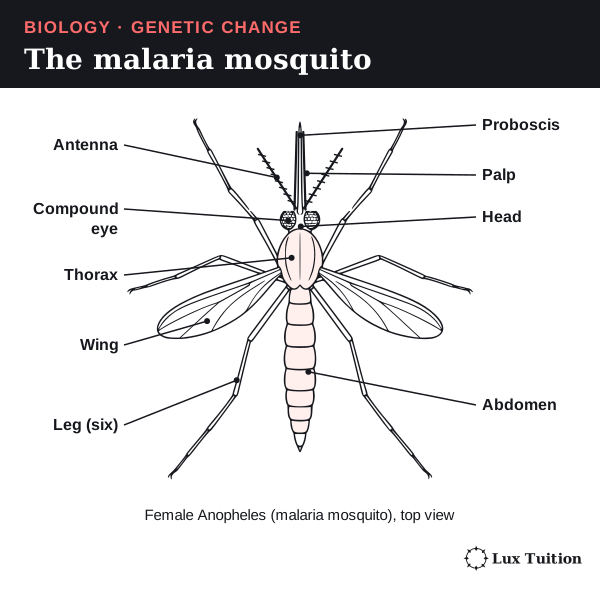
The best-known target is the malaria mosquito (Anopheles). Only females bite, and biting is how they spread malaria between people. A drive that damages a gene needed for female fertility could, in theory, crash a mosquito population.
The figure below labels the main parts. The proboscis is the long, needle-like mouthpart the female uses to feed on blood, with a palp (a short sensory feeler) beside it. The head also carries compound eyes and antennae, the thorax carries the wings and six legs, and the abdomen holds the gut and eggs.
There are real limits. Some mosquitoes carry target-site variants that resist cutting, and the drive can make carriers less likely to survive and breed. Removing a species can affect others in the ecosystem.
Caged laboratory results do not prove a drive would work, or be safe, in the wild, so containment, step-by-step testing and public decision-making are central. Kyrou and colleagues, 2018: laboratory mosquito gene-drive study.
How can genetic research benefit society?
evaluating the potential benefits for society of research using genetic technologies
Genetic research can benefit society before any product is released. Testing what a gene does can reveal how a disease works, failed approaches show which limits are real, and better methods strengthen later studies. Research also has costs: money spent on one project cannot be spent on another, outcomes are uncertain, and benefits may not reach everyone fairly.
How do genetic techniques affect biodiversity?
evaluating the changes to the Earth’s biodiversity due to genetic techniques
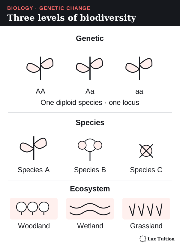
Biodiversity is the variety of life, measured at three levels:
- Genetic diversity: the variety of alleles within a species or population.
- Species diversity: the variety of species in an area.
- Ecosystem diversity: the variety of habitats and communities, such as forests, wetlands and grasslands.
A change at one level does not prove a change at the others: adding a new allele to a crop does not create a new species.
Genetic level
- Can increase: crossing different lines, or adding a sequence the population lacked, adds alleles or combinations.
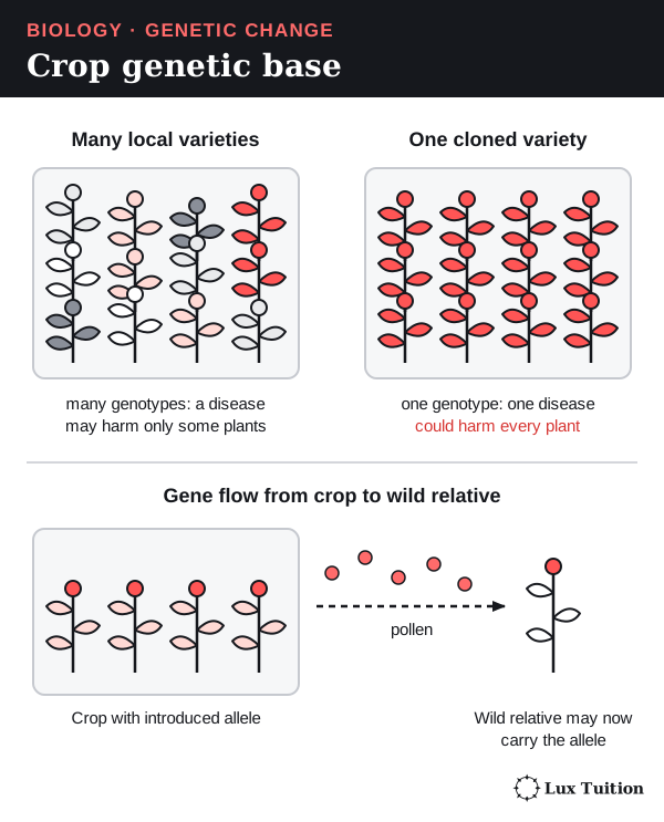
- Can decrease: replacing many local varieties with a few successful ones lowers the total variety across farms. Cloning (making genetically identical copies) of one high-yielding plant adds no new alleles, so a huge crop can rest on a narrow genetic base.
Allelic richness counts how many different alleles exist at the genes studied. Heterozygosity is how often individuals carry two different alleles of a gene. A population can keep both alleles while one becomes very rare, lowering heterozygosity but not richness. Results depend on which genes and populations were sampled.
Species and ecosystem levels
- Using fewer broad-spectrum insecticides can help pollinators and other useful insects.
- Clearing land or changing herbicide use can remove food and shelter for other species.
Not all these effects come from the inserted gene. A fair comparison separates the crop trait from farming practice, location and past land use.
Does artificial manipulation of DNA have the potential to change populations forever?
How do CRISPR genome editing and gene therapy work?
The most widely used genome-editing tool, CRISPR–Cas9, has two parts:
- Cas9, an enzyme that cuts both strands of DNA (a DNA-cutting enzyme is a nuclease);
- a guide RNA, a short RNA matching the target sequence, which steers Cas9 to that one spot.
An edit happens in four steps:
- The guide RNA base-pairs with the matching DNA.
- Cas9 only cuts if a short sequence called the PAM sits right next to the target, which stops it cutting just anywhere.
- Cas9 cuts both strands, making a double-strand break.
- The cell repairs the break, and the repair decides the result:
- End joining glues the ends back, often adding or losing a few bases (an “indel”). This usually switches the gene off.
- Template repair copies a piece of DNA supplied by scientists (a donor template), which can make a planned change.
Results are not guaranteed. They depend on guide design, getting the tool into the right cells, avoiding cuts at similar sequences elsewhere (“off-target” cuts) and unexpected changes at the target.
If only some cells are edited, the organism is a mosaic. Some newer methods change a base without cutting both strands. NHGRI: genome editing.
Gene therapy
Gene therapy treats a disease by adding, changing or controlling genetic material in a patient’s cells. It does not always replace a faulty gene.
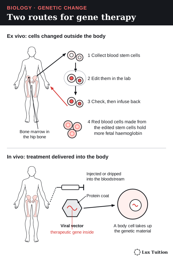
- Ex vivo (“outside the living body”): cells are removed, changed and checked in the lab, then returned to the patient. For blood disorders these are blood stem cells: unspecialised cells in the bone marrow that divide to make all the blood cells.
- In vivo (“inside the living body”): the treatment is delivered straight into the patient, for example by injection.
The carrier that gets genetic material into cells is a vector. A virus is a tiny particle made of genetic material inside a protein coat, which can only multiply inside a living cell. A viral vector is a virus whose disease-causing genes have been removed and replaced with the therapeutic gene. Tiny fat (lipid) particles are another type of vector. Delivery systems differ in capabilities and limitations.
Gene therapy changes somatic cells, so the change is not normally passed to the patient’s children.
Worked example: editing a control switch to treat sickle cell disease
Sickle cell disease is caused by a mutation in the HBB gene, which codes for part of adult haemoglobin, the oxygen-carrying protein in red blood cells. Before birth, a baby makes fetal haemoglobin, which works well. After birth, a gene called BCL11A switches fetal haemoglobin off.
The treatment Casgevy, reviewed by the US FDA in 2023, works like this:
- The patient’s blood stem cells are collected.
- In the lab, CRISPR–Cas9 cuts an enhancer of BCL11A that is only active in developing red blood cells.
- The edited stem cells are returned to the patient (ex vivo therapy).
- As these stem cells make new red blood cells, the developing cells make less BCL11A, so their fetal haemoglobin genes stay switched on.
- The finished red blood cells (which have no nucleus) carry more fetal haemoglobin, which reduces the effects of the faulty adult haemoglobin.
It does not repair the HBB mutation; it edits a non-coding control region. That links non-coding DNA, gene regulation and medicine. It also shows that the molecular result, delivery, patient benefit and long-term safety are separate questions. FDA scientific review, 2023.
Genome-editing mechanisms in diagrams
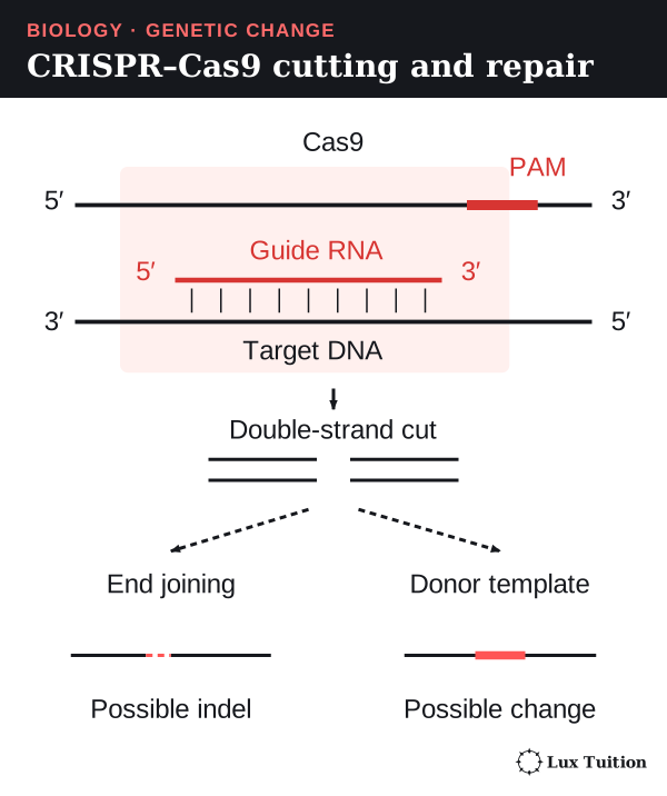
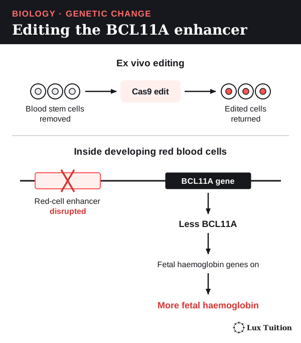
How do artificial insemination and artificial pollination compare?
artificial insemination
artificial pollination
Reproductive technologies are methods where people control how gametes meet. Both technologies here move male gametes to a chosen female, so they support selective breeding. Both still rely on meiosis and fertilisation, so the offspring are not clones: each gets a new mix of alleles from two parents.
Artificial insemination (AI) in livestock
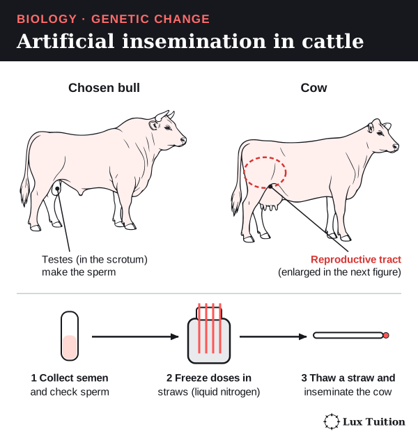
Semen is the fluid that carries sperm. In artificial insemination, people place semen from a chosen male into a female’s reproductive tract instead of the animals mating. In cattle, the male is a bull and the female a cow.
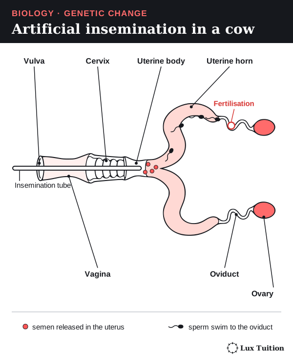
The cow’s reproductive tract, from the outside in, has the vulva (outer opening), vagina, the ringed cervix (a firm “gateway”), the uterus (a short body and two long horns, where the calf develops), two oviducts (where the egg is fertilised) and two ovaries (which release eggs).
The process:
- Collect semen from a selected bull and check it (how many sperm, how well they swim).
- Store it: dilute it, then use it fresh or freeze it in thin straws in liquid nitrogen.
- Time it: watch the cow for signs she is about to release an egg.
- Deposit it: a thin tube is passed through the vagina and guided through the cervix, and the semen is released into the uterine body.
- The sperm swim to the oviduct, where fertilisation happens inside the cow, just as after mating.
Outcomes. One bull’s semen can be split into hundreds of doses and sent around the world, so alleles for valued traits such as high milk yield spread through herds quickly. Screened semen and less animal transport can cut some disease risks, but poor handling can still spread infection or lower pregnancy rates. Costs include trained staff, storage and monitoring.
Effect on diversity. If a few popular bulls father huge numbers of calves, herds become closely related and a hidden harmful recessive allele can spread. Used the other way, AI from under-represented males can help conservation breeding keep diversity.
Artificial pollination
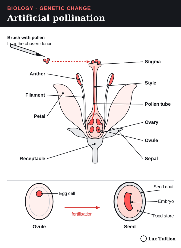
Pollination is the transfer of pollen, which contains a plant’s male gametes, to the female part of a flower. The flower parts involved are:
- the stamen (male part): a pollen-making anther on a stalk called the filament;
- the carpel (female part): a sticky stigma on top, a stalk called the style, and an ovary at the base;
- ovules inside the ovary, each containing an egg cell.
In artificial pollination, a breeder does the transfer by hand:
- Choose the pollen donor and the recipient flower.
- Remove the anthers of the recipient before they ripen, so it cannot pollinate itself.
- Cover the flower with a bag so insects and wind cannot bring other pollen.
- Transfer ripe pollen from the donor’s anthers to the recipient’s stigma, for example with a brush.
- Re-cover and label the flower so the cross can be tracked.
A compatible pollen grain then grows a pollen tube down the style to an ovule and delivers the male gametes, which fertilise the egg cell. The ovule becomes a seed, containing an embryo (a tiny new plant) and a food store inside a protective seed coat. The ovary usually becomes the fruit. Pollination is only the transfer step, not fertilisation.
Outcomes. A breeder can combine, for example, disease resistance from one plant with a quality trait from another, or rescue seed set when insects are scarce. It needs compatible parents and good timing, and it is labour-intensive. Repeated pollination from a narrow set of parents can reduce diversity. The seeds vary because each carries a new combination of the parents’ alleles. University of Minnesota: controlled plant breeding.
Comparing the two
| Comparison | Artificial insemination | Artificial pollination |
|---|---|---|
| What is moved | Semen, containing sperm | Pollen, containing male gametes |
| Where it is placed | Female reproductive tract (usually the uterus) | The stigma |
| What happens next | Sperm swim to the egg; fertilisation may occur | A pollen tube delivers the gametes to the ovule; fertilisation |
| Genetic outcome | Sexual offspring with alleles from both parents | Seeds with alleles from both parents |
| Effect on population | Changes which males father offspring, and how often | Changes which pollen donors father seeds, and how often |
IVF and embryo transfer are related. In in vitro fertilisation (IVF), egg and sperm are joined in a dish and the early embryo is placed in a female’s uterus. Embryo transfer moves an early embryo from one female to another, and can follow IVF or fertilisation inside a donor. Neither makes a clone, and their effect on diversity depends on how many different parents are used.
How does whole-organism cloning work, and how effective is it?
whole organism cloning
A clone is an organism (or cell) made by asexual reproduction, reproduction from a single parent without gametes, so it has the same genotype as its source. Two cautions: new mutations can still arise as a clone grows, and the same genotype does not guarantee the same phenotype, because nutrition, development and gene regulation also matter.
Cloning plants: cuttings and tissue culture
Many plant cells can still become any kind of plant cell and regrow a whole plant, so many plants can be cloned, though success depends on species and culture conditions. It can propagate plants that are difficult to propagate sexually. Some methods multiply meristems (growing tips where cells divide) directly, and clean source tissue, treatment and testing support pathogen-free production.
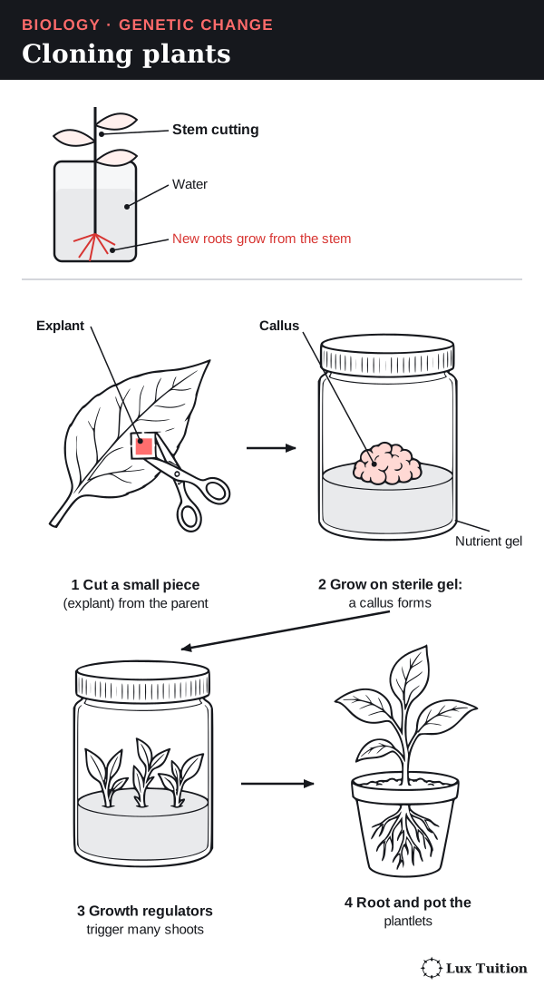
- A cutting is a piece of stem placed in soil or water. Its cells divide by mitosis and differentiate (become specialised) to form new roots. The new plant has the parent’s genotype, apart from any new mutation.
- Tissue culture (micropropagation) clones plants in large numbers. Hormones are chemical signals that control growth; plant hormones used this way are called plant growth regulators.
- A small piece of tissue, the explant, is cut from a leaf of the parent.
- It is sterilised and placed on a sterile gel containing sugar, minerals and growth regulators.
- With some methods it first forms a callus, a lump of unspecialised cells.
- Changing the growth regulators makes shoots, then roots, form. Shoots can be divided again and again.
- The plantlets are moved to soil and gradually acclimatised to normal conditions.
Advantages: thousands of identical plants of a valuable variety (a cultivar) can be made quickly, without the reshuffling of alleles that happens in seeds.
Limits: contamination, cost and unwanted variation that can arise in culture. If a whole region grows one genotype, a disease that attacks one plant can attack them all. Tissue culture does not automatically make plants disease-free.
Cloning animals: embryo splitting
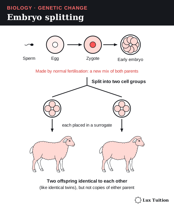
In embryo splitting, an early embryo (a small ball of unspecialised cells) is split into two or more groups. Each is placed into a surrogate mother (a female who carries another animal’s embryo) and can develop into a separate animal.
The animals are clones of each other, like identical twins. But the embryo came from sexual reproduction, so embryo splitting copies a new combination of alleles, not either adult parent.
Cloning animals: somatic cell nuclear transfer (SCNT)
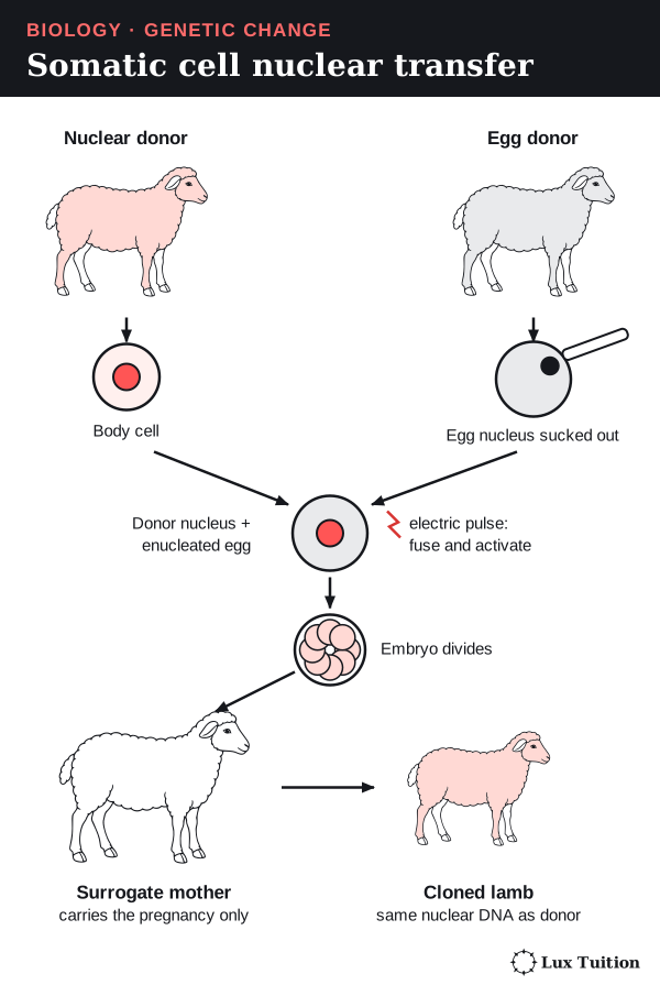
Somatic cell nuclear transfer can copy an adult animal. Dolly the sheep (born 1996) was the first mammal cloned this way from an adult cell.
- A somatic (body) cell is taken from the donor animal to be copied.
- An unfertilised egg cell is taken from a second female, and its nucleus is sucked out with a fine glass needle. This enucleated egg has no nuclear chromosomes but keeps its cytoplasm.
- The donor cell’s nucleus is put into the enucleated egg, or the two cells are fused with a small electric pulse.
- The new cell is activated and starts dividing. Chemicals in the egg’s cytoplasm must reprogramme the adult nucleus to behave like a new embryo’s.
- The early embryo is placed in the uterus of a surrogate mother.
There is no fertilisation in SCNT. Three roles are involved (one animal can sometimes fill two):
- the nuclear donor supplies the chromosomes, so the clone is a copy of this animal’s nuclear genotype;
- the egg donor supplies the cytoplasm, including most of the mitochondrial DNA (the small amount of DNA inside mitochondria), and normally no inherited nuclear DNA;
- the surrogate carries the pregnancy but passes on none of her DNA. NHGRI: cloning processes and limitations.
Injuries are not copied: losing an ear does not erase the ear-development genes, because an unaffected donor nucleus still holds them. But a mutation in the donor nucleus is copied, even if it began as a somatic mutation, which normal inheritance would never pass on.
How effective is cloning?
“Effective” needs a clear finish line: a dividing reconstructed egg is not a healthy adult that can breed. In SCNT, reprogramming is often incomplete, many embryos are lost and pregnancies can have developmental problems (including placental abnormalities), and success varies by species and method, which also affects the welfare of the egg donors and surrogates.
A success rate means nothing unless you know what it is a percentage of: reconstructed eggs, transferred embryos, pregnancies or live births.
Cloning can preserve a valuable genotype and may help some conservation programmes when viable material, reproductive compatibility and healthy development are available. But copying the last few individuals of a species cannot bring back alleles already lost, so making more animals is not the same as restoring genetic diversity.
How does gene cloning use recombinant DNA?
gene cloning
Gene cloning means making many identical copies of one chosen piece of DNA, usually a gene. The product is a collection of DNA molecules, not a new animal or plant.
Start here: inside a bacterium
A bacterium has a rigid cell wall outside its cell membrane. Its main DNA is one large loop, the bacterial chromosome.
Many bacteria also carry plasmids: small, separate rings of DNA that copy themselves independently. Bacteria divide fast and copy their plasmids each time, so a plasmid makes an ideal carrier, or vector, for a gene.
- Recombinant DNA does not have to combine different species.
- A transgenic organism contains DNA from a different species. An organism whose own gene has been edited may contain no foreign DNA, so not every genetically modified organism is transgenic.
A plasmid vector has three key features:
- an origin of replication, where copying starts;
- a site where DNA can be inserted;
- a selectable marker, usually a gene for resistance to an antibiotic (a drug that kills bacteria), so cells carrying the plasmid can be picked out.
To make a protein, an expression vector also needs a promoter that works in the host.
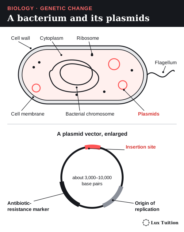
The steps of gene cloning
- Get the gene. It can be cut from a genome (an organism’s complete set of DNA), made artificially or copied by PCR. Ordinary bacterial systems cannot remove human introns, so for a human protein scientists use an intron-free copy. The enzyme reverse transcriptase can build this from mature mRNA; the copy is called complementary DNA (cDNA).
- Choose a restriction enzyme. A restriction enzyme recognises one short DNA sequence, its restriction site, and cuts the backbone there.
- Cut the gene and the plasmid so their ends are compatible (using the same enzyme is one option). Many enzymes cut the two strands at slightly different points. This leaves short single-stranded tails called sticky ends, which match on both pieces. (Some enzymes cut straight across, leaving blunt ends.)
- Join them. The sticky ends base-pair, then DNA ligase seals the backbone with phosphodiester bonds (the bonds linking one nucleotide to the next).
- Put the plasmid into bacteria (transformation), using a brief heat shock or an electric pulse (electroporation). Many cells take up nothing, and some take up a plasmid without the gene.
- Select and check. An antibiotic kills cells without a plasmid. Screening and DNA sequencing then confirm the right gene, the right way round, with no errors.
- Grow the chosen clone. It multiplies, copying the plasmid each time. If a protein is wanted, the protein is then collected and purified.
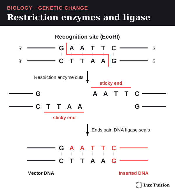
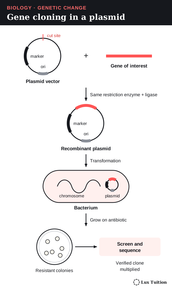
Selection is not proof
A cell with the resistance marker grows on antibiotic whether or not its plasmid holds the gene. Blue–white screening adds a check. Each colony (a visible clump of bacteria grown from one cell) is tested using the gene lacZ. It sits around the insertion site and makes an enzyme that turns a chemical in the gel blue:
- Blue colony: lacZ is intact, so there is usually no insert.
- White colony: lacZ may have been broken by an insert, so the colony may contain the gene.
White colonies are candidates; sequencing checks whether they hold the correct, error-free gene. Addgene: blue–white screening.
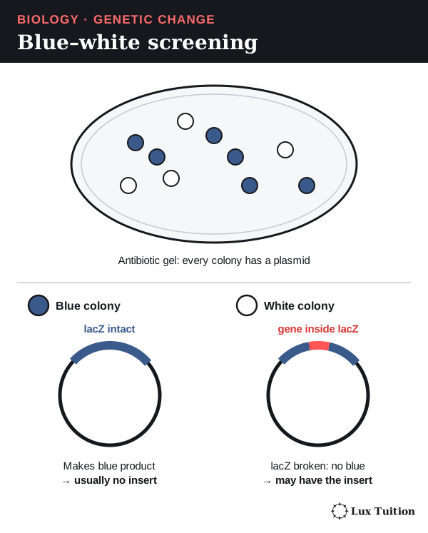
Why bacteria can read a human gene, and why it can still fail
The genetic code is almost universal: nearly all organisms read each codon as the same amino acid, so a bacterium builds the same amino acid chain from a human gene. But the host needs a promoter and translation signals it recognises, and some human proteins need folding or added sugar groups that bacteria cannot provide; yeast or mammal cells suit those better.
How effective is gene cloning? It is good for keeping a checked sequence, studying genes and making products. Contamination, copying errors, unstable inserts, toxic products and poor expression reduce success. It is judged by confirmed DNA identity, copy number and, where relevant, protein activity and purity, not by healthy offspring as in whole-organism cloning.
How do PCR, gels and sequencing support genetic technologies?
Three laboratory tools appear throughout this module, and each answers a different question.
PCR: copying one piece of DNA
PCR makes millions of copies of one chosen section of DNA in a tube, with no living cells. The tube contains:
- template DNA, the sample containing the section to copy;
- two primers, short single strands of DNA that match the two ends of the target, one on each strand;
- heat-stable DNA polymerase, such as Taq polymerase from a hot-spring bacterium;
- free nucleotides (A, T, C and G) and a buffer solution (a liquid that keeps the conditions stable).
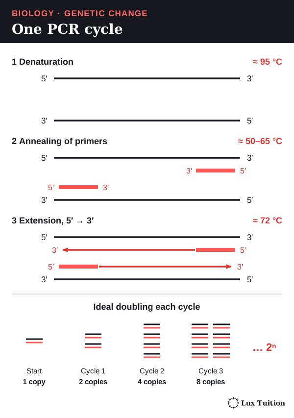
A machine heats and cools the tube in repeated cycles of three steps:
- Denaturation (about 95°C): heat breaks the weak hydrogen bonds holding the base pairs together, separating the strands. (In cells, an enzyme called helicase does this.)
- Annealing (about 50–65°C, depending on the primers): cooling lets the primers bind to their matching sequences.
- Extension (about 72°C for Taq): polymerase adds nucleotides to the 3′ end of each primer, building new strands 5′ → 3′.
The new strands become templates in the next cycle, so the target ideally doubles every cycle.
Worked calculation. With perfect doubling, N = N₀ × 2ⁿ (N₀ = starting copies, n = cycles). Ten molecules after 10 cycles give 10 × 2¹⁰ = 10,240 copies. Real reactions slow and level off, so this is a model, not a guaranteed yield.
PCR only copies DNA; it does not put a gene into an organism or make a protein. Copying errors and contamination mean products may need checking. NHGRI: PCR.
Gel electrophoresis: sorting DNA by size
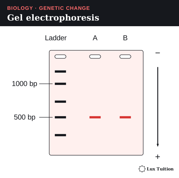
Gel electrophoresis separates pieces of DNA by length.
- Samples are loaded into wells at one end of an agarose gel, a jelly with tiny pores.
- An electric current is applied. DNA is negatively charged (from the phosphates in its backbone), so it moves towards the positive electrode.
- Small pieces slip through the pores more easily, so they travel further in the same time.
- A DNA ladder of known sizes runs in one lane as a ruler, and stain makes the bands visible.
A band’s position estimates its size (for comparable linear fragments; uncut plasmid forms migrate differently), and its brightness roughly shows the amount. But pieces of equal length can have different sequences, so a band at the expected size supports but does not prove identity.
Sequencing, probes and controls
DNA sequencing reads the exact base order, so it can confirm an inserted gene, find a mutation or check an edit. A DNA probe is a short, labelled single strand that sticks to its complementary sequence, showing where that sequence is.
- PCR asks: can this region be copied from the sample?
- A gel asks: how long are the pieces?
- Sequencing asks: what is the exact base order?
Controls separate a failed test from a truly absent target. A positive control contains the target and should work. A no-template control has no DNA added; a band there means contamination. A clean negative control cannot prove that DNA was extracted from your sample.
How does Bt cotton work as a transgenic crop?
the development of transgenic organisms in agricultural and medical applications (ACSBL087)
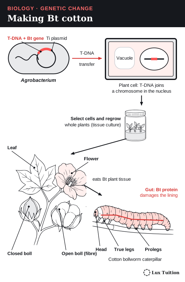
Bt cotton is a transgenic crop carrying genes from a soil bacterium, Bacillus thuringiensis (“Bt”). These genes let the plant make its own insecticidal proteins, which kill certain caterpillar pests, such as the cotton bollworm, that eat the leaves and bolls (the seed pods that hold the fibre). It is not cotton sprayed with bacteria, and it does not kill all insects.
How Bt cotton is made
One method uses another soil bacterium, Agrobacterium, which naturally transfers part of its DNA into plant cells. Scientists use a “disarmed” version that causes no disease.
- Build the construct. The Bt gene, a plant promoter and a selectable marker are placed in the transferable region, the T-DNA, of the bacterium’s Ti plasmid.
- Transfer. Agrobacterium moves only the T-DNA into cotton cells, where it can join a plant chromosome.
- Select. Only cells carrying the marker survive on the growth medium.
- Regenerate whole plants from these cells by tissue culture.
- Check and breed. Tests confirm the gene is inserted and switched on, and breeding trials check it is stably inherited.
Another method fires DNA-coated particles into cells (particle bombardment).
How the Bt protein works
- A caterpillar eats leaf or boll tissue containing the Bt protein.
- In the caterpillar’s gut, the protein is activated and binds to specific targets on the gut lining.
- The gut wall is damaged, so the caterpillar stops feeding and dies.
Because the targets are specific, only susceptible insect groups are affected. Less feeding damage protects the crop and can reduce some insecticide spraying. Insect resistance is different from herbicide tolerance (surviving a weedkiller); a crop can carry either trait or both. Australian OGTR: GM cotton.
Resistance and evaluation
Resistance can evolve. Caterpillars carrying alleles that let them survive Bt protein breed while the others die, so by natural selection the resistance allele becomes more common. Two strategies slow this:
- Stacking two or more Bt proteins in one plant, so an insect needs resistance to all of them;
- Refuges: areas of non-Bt crop where susceptible insects survive and mate with resistant ones, keeping resistance alleles rare. Both work only conditionally: resistance genetics, mating and management decide how well.
Neither makes evolution impossible.
A fair evaluation compares crop damage, yield, spraying, costs and effects on other organisms with a suitable non-Bt system. Bt cotton can improve pest control, but its long-term value depends on managing resistance and on local ecology and economics.
What are the benefits of recombinant DNA in medicine and industry?
Medicine: recombinant human insulin
Insulin is a hormone that lowers blood sugar. People with type 1 diabetes cannot make enough and must inject it; before recombinant insulin, it was extracted from animals.
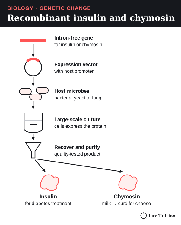
- An intron-free insulin gene (or one for a precursor later trimmed into insulin) is placed in an expression vector.
- The vector is put into a host: bacteria or yeast.
- The host cells are grown in huge tanks, where they make the protein.
- The protein is collected, folded correctly and purified.
Patients receive only the purified insulin, not the microbes.
Benefits: a reliable supply of a human protein, less dependence on animal sources. Limits: quality control is still needed, and price and distribution decide who can get it. It was approved in the USA in 1982; modern products are not all made the same way. FDA: the history of insulin.
Research: genetically modified animals
A transgenic animal is made in four steps:
- Build the DNA construct: the chosen gene plus a promoter, prepared as in gene cloning.
- Microinject it, using a fine glass needle, into a fertilised egg or very early embryo (in mosquito research, into eggs). Because this happens so early, the cells formed by later divisions, ideally including the germ line, carry the gene.
- Screen the offspring with PCR or sequencing to find those carrying the gene, and test that they make the protein.
- Breed the animals that pass the gene to their own offspring, to establish a transgenic line.
Comparing animals with one gene switched off (“knocked out”) against normal animals can reveal that gene’s role, and adding a disease-causing allele can model part of a human disease. Results must allow for genetic background, environment and unintended changes, and they do not automatically apply to humans.
Benefits are weighed against welfare using the “3Rs”: replace animals where possible, reduce numbers and refine procedures to reduce suffering.
Industry: recombinant chymosin
Chymosin is a cheese-making enzyme. It cuts a milk protein called κ-casein, which destabilises casein micelles (clumps of milk protein) so milk proteins clump into solid curds. It used to be extracted from animals; now the bovine chymosin gene is expressed in a production microorganism (such as GM Trichoderma reesei) and the enzyme is purified.
In its 2022 assessment A1244, Food Standards Australia New Zealand assessed chymosin made this way. FSANZ A1244 approval report.
Benefits: a consistent supply that does not depend on animals. Limits: fermentation, purification and quality checks cost money, enzyme properties must be controlled and the production organism contained. A purified enzyme and a living GM organism are different products with different risks. Whether a cheese suits a diet or culture depends on the actual production process and community or certification advice, not the word “recombinant”.
| Application | How it delivers the benefit | Evidence needed for a balanced judgement |
|---|---|---|
| Bt cotton | The plant makes proteins that cut damage by target pests. | Local pests, resistance, spraying, effects on other species, yield, cost. |
| Recombinant insulin | Host cells make a human protein for purification. | Activity, purity, supply, cost and patient access. |
| Recombinant chymosin | A microbe makes a milk-clotting enzyme. | Performance, purity, efficiency and comparison with other sources. |
“Genetic technology” is too broad for one verdict: the organism, gene, product, exposure and purpose decide which evidence matters.
How does agricultural biotechnology affect biodiversity?
Agricultural biotechnology can change biodiversity in both directions, through four main pathways.
- Narrowing the genetic base (above): one cloned genotype everywhere means a disease that attacks it could attack every plant.
- Adding variation by crossing lines or adding a gene.
- Gene flow to other populations. Pollen or seed can carry an introduced allele into nearby compatible plants, including wild relatives. Whether it persists depends on reproduction and selection: a pesticide-resistance allele may help only where that pesticide is used, and resistance can carry costs elsewhere. Gene flow is a possibility to investigate, not proof that every modified crop becomes invasive.
- Changes to farm ecosystems (above): fewer broad-spectrum sprays can help other insects; land clearing can harm them.
Biotechnology can also protect biodiversity:
- Seed banks and gamete banks store samples of genetic variation.
- DNA testing shows how closely animals are related, so breeders avoid pairing close relatives.
- Assisted reproduction gives under-represented founders more offspring.
- Cloning stored cells might restore a lost genotype, but cannot recreate variation that was never stored.
These tools support, but do not replace, protecting habitat.
For exam judgements, follow the chain: technique → change in reproduction or phenotype → change in which alleles are passed on → measured genetic or ecological effect. Different breeding choices, environments and time scales give different results.
How do social, economic and cultural contexts influence genetic technologies?
Whether a biotechnology is used depends on more than the science. Social context is what a community values and fears, economic context is cost and who can pay, and cultural context is beliefs, traditions and identity.
Human genetic technologies. A person’s genetic data also reveals information about their relatives, so consent, privacy, how long samples are kept and who can access follow-up data matter. Somatic therapy treats one patient, but in heritable editing future descendants cannot consent to the change they inherit. High prices and specialist hospitals can limit access even when a treatment works.
Cultural views can shape attitudes to embryos, animal products, family, ownership of biological material and responsibilities to land and other species. Views differ even within one community, so good consultation involves the affected people, including appropriate Indigenous governance where biological material, knowledge or Country is involved. It is not sound to assume one opinion for a whole cultural group.
Worked judgement: human genome editing. The World Health Organization’s 2021 recommendations came from a two-year consultation with hundreds of participants, including scientists, patient groups, faith leaders and Indigenous peoples. It recommended oversight in nine areas, including trial registries, illegal or unsafe research, and “education, engagement and empowerment”, and stressed benefits “for all people”. WHO: human genome-editing recommendations, 2021.
Judgement: social and cultural context shaped the governance itself, adding public engagement, monitoring and fairness to the technical safety rules. The source shows what was recommended, not how any one community feels about a particular use.
Interpreting different sources. Each answers a different question:
- the OGTR cotton fact sheet explains one crop in the Australian regulatory setting;
- the FSANZ chymosin report assesses one enzyme application;
- the FDA Casgevy review covers one medical product;
- WHO considers broad governance.
Worked judgement: Casgevy. The FDA review shows the sickle cell treatment works by editing a patient’s own blood stem cells. The process needs cell collection, chemotherapy to make room for the edited cells, and a long stay in a specialist hospital. The UK health-cost body NICE lists the treatment at £1,651,000 per patient before discounts. NICE TA1044.
Judgement: the science works, but the economic context (who pays) and specialist facilities limit who can get it more than the biology does. These sources cannot show how communities feel about the treatment; that would need a survey or consultation source.
A decision about one product is not approval of all biotechnology. A farmer’s costs or a community statement add perspectives but answer different questions from an experiment.
Working scientifically with genetic-change evidence
Being precise about what was actually measured prevents most errors; the methods are in the Working Scientifically skills.
Worked comparison 1: crop damage in a field trial
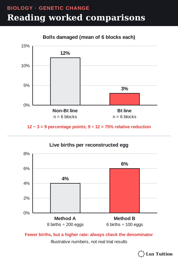
An illustrative trial compares a Bt cotton line with a closely related non-Bt line in six randomly assigned field blocks, with water, planting density and management kept the same.
- Non-Bt line: 12% of bolls damaged (mean).
- Bt line: 3% of bolls damaged (mean).
- Absolute difference: 12 − 3 = 9 percentage points.
- Relative reduction: (12 − 3) ÷ 12 × 100 = 75% compared with the non-Bt line.
These numbers are invented to show the method.
What the result does and does not show. The measured result is less crop damage; that is consistent with Bt protein reducing feeding by susceptible pests, a mechanism shown by separate laboratory studies. But two means do not show whether the difference is statistically significant (unlikely to be due to chance), and they say nothing about yield or safety. To judge the evidence, consider:
- block-to-block variation, pest identification, sample size and measuring method;
- the unit of replication: treating every boll on one plant as a separate result exaggerates the evidence;
- repeating across sites and seasons, with suitable comparison lines and “blind” scoring (the scorer does not know which group each sample is from);
- conditions: under low pest pressure the result might differ, so predictions should be narrow, not “Bt always improves yield”.
Worked comparison 2: cloning success
Illustrative method A gives 8 live births from 200 reconstructed eggs: 8 ÷ 200 = 4%. Method B gives 6 from 100: 6%. B has the higher rate despite fewer births.
Neither number shows which method is better for healthy adult survival, cost or welfare. A fair comparison needs similar species, donors, transfer conditions, follow-up and the developmental stages at which losses occur, and small samples leave uncertainty (the range the true value probably lies in). A graph should show sample sizes and define the endpoint (here, live births per reconstructed egg).
Worked comparison 3: judging sources
- A regulatory assessment gives detailed evidence about the specific product and use it assessed.
- A manufacturer’s summary may explain how a product should work but omit its limits.
- An original study shows methods and uncertainty; a later review places it among other studies.
- Newer is not always better: an original report may be best for a discovery date, a current assessment for present performance.
Safety, ethics and communication
Genetic material and microbial cultures need proper containment and disposal. Students must not be exposed to mutagens such as UV: use secondary data or safe models, and keep any UV demonstration enclosed, shielded and teacher-run. Human samples need consent and privacy, and animal work needs welfare safeguards.
Clear communication keeps evidence and prediction separate:
- a sequence diagram shows strand direction and whether the bases are DNA or RNA;
- an allele-frequency calculation states the population size and the number of chromosome sets (ploidy);
- a graph labels units and uncertainty;
- an evaluation ties its judgement to one named application.
Peer feedback can catch a missing control or an over-general claim.
So, can artificial manipulation change populations forever?
Changes last when altered DNA is inherited, or when breeding repeatedly shifts allele frequencies. But selection, drift, gene flow and changing environments keep acting, so permanence is not guaranteed. A somatic edit stays in one person; a germ-line change can enter later generations.
Working Scientifically skills for Module 6
These skills earn marks in every Module 6 question that gives you data, a method or a source.
Questions and hypotheses
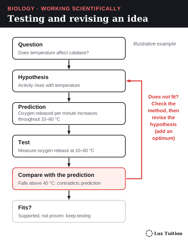
A scientific question asks something data could answer. A hypothesis is a testable explanation, and a prediction says what you should observe if it is right. A testable hypothesis names what you change and what you measure, gives a range, and could be shown wrong. A directional or comparative statement can be a hypothesis; adding a “because” reason is a strong scaffold, not a requirement.
Module 6 example (a safe model). Genetic drift can be modelled with beads: a bag of 50 red and 50 white beads is the gene pool, and each draw is one generation’s breeding sample.
- Hypothesis: allele frequencies drift further from 0.5 in small populations, because each generation is a smaller chance sample.
- Prediction: across 10 trials, samples of 10 beads will show a wider range of red-bead frequencies than samples of 50.
Modifying a hypothesis. If both sample sizes vary about the same, first check the method: three trials may be too few to see the pattern. If more trials still show no difference, narrow or rewrite the hypothesis and test it again with new data.
Common mistake: giving a bare prediction (“more variation”) with no testable link between the variables when a hypothesis is asked for.
Variables, controls and replication
- The independent variable (IV) is what you deliberately change; the dependent variable (DV) is what you measure.
- Controlled variables are kept the same in every group so only the IV can explain a difference.
- An experimental control is an extra group treated identically except for the factor being tested, so you can see what happens without it.
- Biological replication means several independent units per group; repeated measurements of one unit check the measuring, not the biology.
Module 6 example: a Bt cotton field trial. IV: cotton line (Bt or non-Bt). DV: percentage of bolls damaged. Controlled variables: site, season, water, planting density and pest pressure. The experimental control is a closely related non-Bt line grown the same way.
Replicates are separate field blocks; the worked crop-damage comparison shows how to read the results.
Common mistake: calling the controlled variables “the control”. In PCR, the no-template control is a true experimental control: a tube with no DNA that should show no band (see controls).
Risks, ethics, materials and technology
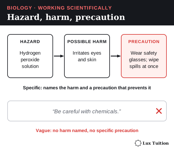
A risk assessment links hazard → possible harm → precaution. “Be careful” earns nothing.
| Hazard | Possible harm | Precaution |
|---|---|---|
| UV lamp (mutagen) | Burns to eyes and skin; DNA damage | Students are not exposed; any demonstration is enclosed, shielded and teacher-run |
| Gel electrophoresis tank | Electric shock | Lid closed while running; switch off before touching the gel |
| DNA stain | Some stains are mutagens | Use a safer stain; gloves; approved disposal |
| Bacterial or yeast cultures | Infection | Approved harmless strains; sealed plates; approved disposal |
Ethics asks whether a study is acceptable, not just safe: genetic data need consent and privacy, and GM animal research needs ethics approval and the fewest animals possible. Materials and technology must be justified: a micropipette that measures 1 µL steps is chosen to load small, equal volumes into gel wells.
Accuracy, precision, reliability and validity
- Validity: the investigation answers the question it claims to. A controlled experiment (only the IV changes) is one way to achieve this. An observational study can also be valid if exposure is measured accurately, groups are matched or adjusted for confounders (other factors linked to both the exposure and the outcome), outcomes are measured consistently and follow-up is long enough. A band at the right size on a gel does not validly show the right gene, because size is not sequence; sequencing does (see gels).
- Reliability: repeating the investigation gives consistent results, such as the same band sizes on three separate gels. Natural variation between organisms is not poor reliability.
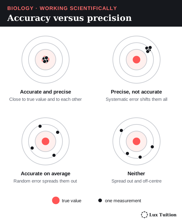
- Accuracy: how close a value is to the true value, such as a band estimated at 510 bp when the fragment is truly 500 bp (“bp” = base pairs).
- Precision: how close repeated values are to each other.
Calibration means checking an instrument against a known standard. A micropipette can be checked by weighing the water it delivers (1 µL of water has a mass of about 1 mg).
Resolution is the smallest change an instrument can show; a pipette adjustable only in 1 µL steps cannot directly set volumes differing by 0.5 µL. Check its working range, adjustment increment and measurement uncertainty separately.
Random error scatters values both ways and lowers precision; averaging independent repeats reduces its effect. Systematic error shifts values in a consistent way, either by a fixed offset or in proportion (for example, always 5% high), and lowers accuracy; a mislabelled ladder would make every size estimate wrong. Averaging cannot remove that bias; calibration or a corrected method can. Uncertainty is the range the true value probably lies in, written as 500 ± 20 bp.
Values written as mean ± standard deviation (± means plus or minus) show spread, the typical distance of values from the mean. That is a different idea from uncertainty in the mean. Overlapping ranges alone do not make a difference unconvincing: sample size and the spread of the mean matter.
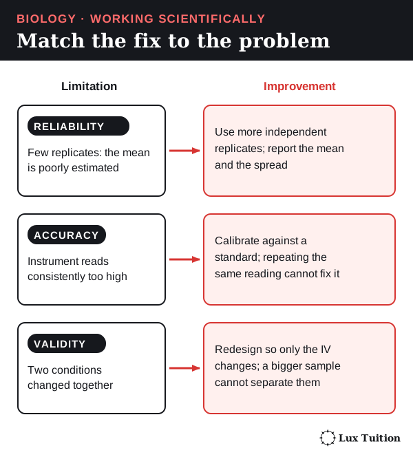
Evaluating and improving. Name a limitation in this investigation, the quality it affects, and a specific fix. Example: “Only one field block per line was used, so local soil differences could explain the result (validity and reliability). Use several randomly placed blocks per line and compare means and ranges.”
Common mistake: expecting repeats to fix a biased instrument. Calibration removes systematic bias; independent repeats improve reliability and the estimate of the mean.
Graphs, best fit, interpolation and extrapolation
Choose the graph by the IV. A categorical variable sorts data into groups (such as the type of biotechnology), so use a column graph. A continuous variable can take any value (such as maternal age), so use a scatter or line graph.
Draw it step by step (HSC exams ask for this, for example a graph of birth-defect prevalence against maternal age):
- IV on the x-axis, DV on the y-axis.
- Label each axis with quantity and unit: “Maternal age (years)”.
- Choose an even scale that fills most of the grid.
- Plot each point as a small cross; use a key if there is more than one data series.
- Draw a line or curve of best fit that follows the trend with points balanced on both sides. Do not join the dots when drawing a trend line or making predictions; for a recorded time series, joining successive observations is fine. Follow the question’s instruction.
Interpolation reads a value inside the measured range; extrapolation extends the trend beyond it. To find when the trend reaches a threshold, draw a construction line across from the threshold to the curve, then down to the x-axis, and leave the lines visible. Extrapolated answers are less certain, because nothing was measured there.
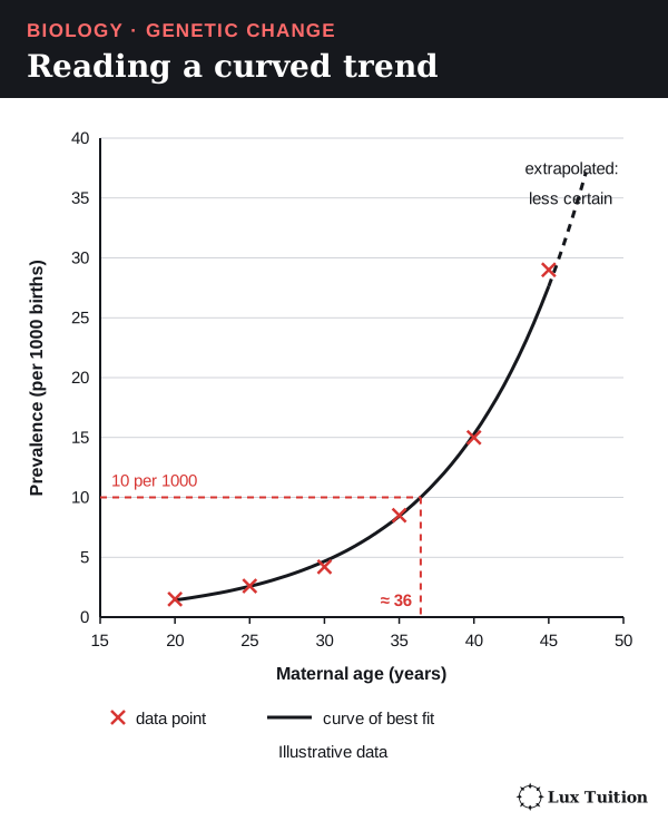
To describe this trend, give direction and values: prevalence rises slowly from 20 to 30 years, then much faster after 35. The rate of increase itself increases with age.
Calculations
- Mean = sum ÷ number of values; median = order the values, then take the middle value, or average the two middle values when there is an even number; range = largest − smallest. Always report a mean with its sample size and spread.
- Percentage change = (new − old) ÷ old × 100. Percentage points are the plain difference between two percentages (see the crop-damage example).
- Rates for different-sized groups: prevalence of 8 per 1000 births equals 8 ÷ 1000 × 100,000 = 800 per 100,000.
- Percentage to a count: if 75% of 40,000 hectares is Bt cotton, that is 0.75 × 40,000 = 30,000 hectares.
Module 6 example (illustrative). Transgenic salmon in a trial reach market size in 18 months; non-transgenic salmon take 30 months. The time saved is (30 − 18) ÷ 30 × 100 = 40% less time. Always state the denominator: here, the non-transgenic time.
For allele frequencies and PCR doubling, see the worked allele-frequency and PCR calculations.
Common mistake: confusing percentage points with percentage change.
Trends, causes and models
Describe the pattern, explain it with a mechanism, and evaluate how well the evidence supports the explanation. An association (two things changing together) is not proof of cause: a confounding variable is another factor linked to both.
Module 6 example. National cotton yield rose as the percentage of Bt cotton grown rose. Bt protein reducing bollworm damage is a plausible cause, but weather, irrigation, fertiliser and other pests also changed over those years. A stronger claim needs side-by-side Bt and non-Bt fields in the same seasons.
A model is a simplified representation with assumptions. The bead model of drift assumes random breeding and no selection or migration; the PCR equation N = N₀ × 2ⁿ assumes perfect doubling. State what a model explains and where its assumptions fail.
Secondary sources and referencing
A secondary source reports data or ideas collected by someone else. Check who wrote it, where it was published, when, what evidence it shows, why it was written and whether it fits your question. Independent corroboration means separate studies with their own data agree. Turning source findings into a judgement is modelled in the Casgevy and WHO worked judgements.
Reference each source consistently, beside the claim it supports, for example: Food Standards Australia New Zealand (2022). Approval report – Application A1244. FSANZ. URL, accessed date.
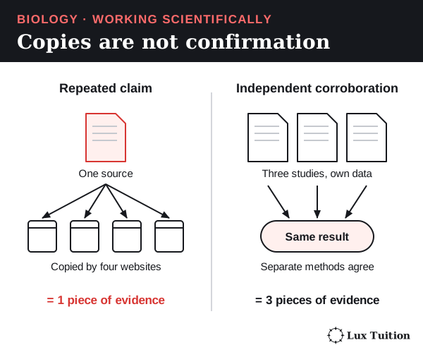
Evidence-based arguments and responding to criticism
Build each argument from four parts: claim, evidence (with values), reasoning (the biology) and limitation.
Module 6 example. “Bt cotton reduced pest damage (claim): damaged bolls fell from 12% to 3% in the illustrative trial (evidence). This is consistent with Bt protein harming the gut of susceptible caterpillars, shown in separate laboratory studies (reasoning). One season was tested, so resistance and other pests are not yet assessed (limitation).”
Responding to criticism: answer each point with evidence. If a critic says the drop was due to good weather, point to the side-by-side comparison in the same season, or collect data across more seasons.
Command words
| Command word | What the answer must do | Answer shape |
|---|---|---|
| Identify | Name | One precise term |
| Describe | Give features, with values | What happens; no reasons needed |
| Explain | Show how or why | Cause → mechanism → consequence |
| Compare | Similarities and differences | “Both… However, X… whereas Y…” |
| Assess / Evaluate | Judge using criteria | Strengths and limitations, then a judgement |
| Justify | Support a claim with evidence | Claim, then evidence and reasons |
| To what extent | Judge how far the evidence supports a claim | Support, limits, then “to a large/limited extent” |
| Analyse | Break into parts; show relationships/trends | Parts or trends, with evidence for each |
| Discuss | Weigh considerations on more than one side | Considerations, evidence, implications, conclusion |
Module 6 example: “To what extent do the data support using Bt cotton for pest control?” Show where the data support it (yield up, bollworm spraying down), where they do not (spraying for other pests may rise, resistance can evolve), then finish with a judgement tied to the data.
These shapes are guides, not formulas: what a question needs depends on its wording and evidence (a justified rejection needs no invented advantage; “compare” needs both similarities and differences). See the NESA glossary. Common mistake: describing when asked to explain, or listing points with no final judgement in “evaluate”, “assess” or “to what extent”.
Where do these ideas connect across Year 12 Biology?
Module 5 explains normal inheritance and protein synthesis in more depth: see Module 5: Heredity. The other Year 12 notes are:
Return to the HSC Biology notes overview for course navigation.
Biology tutoring in Blacktown
Our Year 12 Biology classes in Blacktown walk through these ideas step by step. Students get feedback on how they use them in exam answers. Classes have 1–5 students. See current pricing or enquire about a free trial.
Learn it properly with a specialist tutor at Blacktown or online across NSW. Enquire about a free trial — no cost, no obligation. See online formats and class sizes.
Realistic study tips to help you prepare better for assessments with less stress, plus a printable study planner